Knee Muscle Anatomy Mri : Mri Knee Anatomy Knee Sagittal Anatomy Free Cross Sectional Anatomy : General anatomy and musculoskeletal system.. Knee anatomy wolfgang fitz, md jeffrey lange, md dr. Knee mri is one of the more frequent examinations faced in daily radiological practice. Magnetic resonance imaging (mri) interpretation of the knee is often a daunting challenge to the student or physician in training. This mri knee cross sectional anatomy tool is absolutely free to use. Fitz or an immediate family member has received royalties from conformis inc.;
Master leg and knee anatomy using our topic page. Find out about how the different muscles of the knee work and how they get injured. This long muscle flexes the knee. The semimembranosus muscle is the largest of the posteromedial muscles continuing inferiorly to this level. This mri knee cross sectional anatomy tool is absolutely free to use.
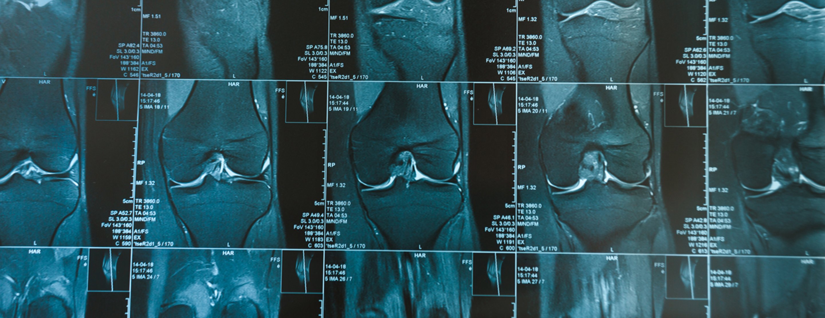
How To Read The Normal Knee Mri Kenhub from thumbor.kenhub.com Musculoskeletal radiology south texas radiology group. In relation to the pcl, the ligament of humphrey courses anterior, and the ligament of wrisberg courses posterior. Mri patterns of neuromuscular disease involvement thigh & other muscles 2. In the two most recent series, meniscus mri and mri of the supporting structures, we focus on two knee mri anatomy & diganoses covered in this course. Want to learn more about it? The main knee muscles are the quadriceps, hamstrings and calf muscles. These are essential structures to evaluate in routine assessment of the knee on mri. Learn anatomy using a full pacs!
General anatomy and musculoskeletal system.
Learn about the muscles, tendons, bones, and ligaments that comprise the knee joint anatomy. Anatomy basic knee mri checklist. Involved early gray = muscle: Any tightness or weakness in the muscles around the knee makes you prone. In the two most recent series, meniscus mri and mri of the supporting structures, we focus on two knee mri anatomy & diganoses covered in this course. To begin, we use a coronal scan of a left knee. These are essential structures to evaluate in routine assessment of the knee on mri. If the knee is flexed more than 5 degrees, it may appear lax. Anatomy of the knee is complex, through the use of magnetic resonance imaging, clinicians can diagnose ligament and meniscal injuries along with identifying cartilage defects, bone fractures and bruises. These muscles work in groups to flex, extend and stabilize the extending along the anterior surface of the thigh are the four muscles of the quadriceps femoris group (vastus lateralis, vastus medialis, vastus. Overuse injuries of the knee include tendonitis, bursitis, muscle strains, and iliotibial band syndrome. Involved early gray = muscle: Musculoskeletal radiology south texas radiology group.
Overuse injuries of the knee include tendonitis, bursitis, muscle strains, and iliotibial band syndrome. The muscles of the knee include the quadriceps, hamstrings, and the muscles of the calf. And has received research or institutional. The main knee muscles are the quadriceps, hamstrings and calf muscles. Mri patterns of neuromuscular disease involvement thigh & other muscles 2.
Knee Mri Scan from www.ucsfhealth.org Mri patterns of neuromuscular disease involvement thigh & other muscles 2. Involved early gray = muscle: Aberrant and accessory muscles around the knee are best identified with mri. Anatomy of the knee can be complicated and hard to understand. Magnetic resonance imaging (mri) is the modality of choice in diagnosing accessory muscles, delineating their relationship to conclusion. Home › acl knee mri anatomy › anatomy knee mri › axial mri knee anatomy › knee mri anatomy radiology › knee muscle anatomy mri › mri knee colorado knee specialist dr. Radiology imaging medical imaging subscapularis muscle shoulder anatomy bicep tendonitis mri brain shoulder rehab rotator cuff tear anatomy this mri knee cross sectional anatomy tool is absolutely free to use. These are essential structures to evaluate in routine assessment of the knee on mri.
Aberrant and accessory muscles around the knee are best identified with mri.
David rubin and robin smithuis. Knee anatomy the orthopedic sports medicine institute in they. The main knee muscles are the quadriceps, hamstrings and calf muscles. Scroll through the structures to understand the anatomy. Anatomy, symptoms, and radiologic evaluation. Free cross sectional anatomy of the knee based on mri : General anatomy and musculoskeletal system. View of the anatomical labels. If the knee is flexed more than 5 degrees, it may appear lax. Use the checklist to quiz yourself. They move when you do—when you walk, run, dance, stretch your legs, or make any action you can think of that there are two muscle groups that act on the knee joint: Mr arthrogram knee loose osteochondral lesion. Knee muscles need to have both good strength and flexibility.
The muscles of the knee include the quadriceps, hamstrings, and the muscles of the calf. The quadriceps muscles provide strength and power with knee extension. Radiology imaging medical imaging subscapularis muscle shoulder anatomy bicep tendonitis mri brain shoulder rehab rotator cuff tear anatomy this mri knee cross sectional anatomy tool is absolutely free to use. Involved early gray = muscle: To begin, we use a coronal scan of a left knee.
Knee Anatomy Magnetic Resonance Mr Axial from konez.com Radiology imaging medical imaging subscapularis muscle shoulder anatomy bicep tendonitis mri brain shoulder rehab rotator cuff tear anatomy this mri knee cross sectional anatomy tool is absolutely free to use. Scroll through the structures to understand the anatomy. The main knee muscles are the quadriceps, hamstrings and calf muscles. Serves as a paid consultant to or is an employee of conformis inc.; Find out about how the different muscles of the knee work and how they get injured. The journal of musculoskeletal medicine. Anatomy, symptoms, and radiologic evaluation. Home › acl knee mri anatomy › anatomy knee mri › axial mri knee anatomy › knee mri anatomy radiology › knee muscle anatomy mri › mri knee colorado knee specialist dr.
Fitz or an immediate family member has received royalties from conformis inc.;
Involved early gray = muscle: These muscles work in groups to flex, extend and stabilize the extending along the anterior surface of the thigh are the four muscles of the quadriceps femoris group (vastus lateralis, vastus medialis, vastus. General anatomy and musculoskeletal system. Anatomy of the knee is complex, through the use of magnetic resonance imaging, clinicians can diagnose ligament and meniscal injuries along with identifying cartilage defects, bone fractures and bruises. Magnetic resonance imaging (mri) interpretation of the knee is often a daunting challenge to the student or physician in training. This long muscle flexes the knee. Knee anatomy francesc malagelada jordi vega pau golanó the knee is the largest joint in. On anatomical parts the user. Free cross sectional anatomy of the knee based on mri : Want to learn more about it? The muscles of the knee include the quadriceps, hamstrings, and the muscles of the calf. And has received research or institutional. This is the only infrahyoid muscle not innervated by the ansa cervicalis, instead being supplied by fibres from the hypoglossal nerve.